Leukoplakia: Symptoms, Causes and Treatment
Reader-supported. We may earn a commission from links on this page. Advertising disclosure.
In this article
What is Leukoplakia?
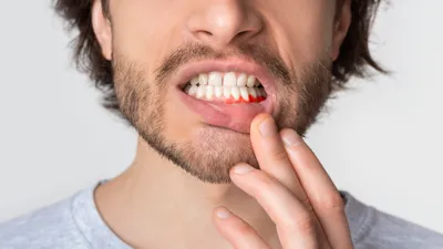
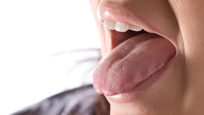
Leukoplakia causes white patches or plaque to form on the gums, tongue, or oral mucosa. It can resemble oral thrush, a yeast infection of the oral cavity.
However, unlike oral thrush, leukoplakia can turn into a mouth cancer. Around 3 to 17.5% of people with leukoplakia develop squamous cell carcinoma, the most common type of oral cancer, within 15 years.
The different types of leukoplakia include:
- Homogenous — White lesions with uniform, smooth, wrinkled, or ridged surfaces.
- Non-homogenous — White or white and red patches with a non-uniform appearance and irregular surface texture.
- Idiopathic — A rare potentially malignant lesion, usually found on the tongue with an increased risk of malignant transformation as compared to the tobacco-associated form.
- Proliferative verrucous leukoplakia (PVL) — A type of non-homogenous leukoplakia causing extensive raised white plaques with a wart-like surface.
- Hairy leukoplakia — White, hairy lesions with a fuzzy appearance. It’s common in people with a weakened immune system due to Epstein-Barr virus (EBV) or HIV.
What Causes Leukoplakia?
The cause of leukoplakia is unknown, but it’s thought to be related to chronic irritation or inflammation of the mucous membranes. Leukoplakia is also strongly associated with tobacco use.
Age also seems to play a role as fewer than 1% of leukoplakia cases occur in people under 30. Meanwhile, most cases occur in men between 50 and 70.
Although it’s not fully understood, there seems to be a link between human papillomavirus (HPV) and oral leukoplakia. Some research shows a strong association between HPV infection and leukoplakia. Lastly, it can also happen because of wearing ill-fitting dentures or having broken or sharp teeth that rub against the cheeks or tongue.
Risk Factors for Leukoplakia
Chewing and smoking tobacco are significant risk factors for developing leukoplakia. This includes other forms of smokeless tobacco such as snuff, which are finely ground tobacco leaves that are inhaled through the nose.
In some parts of the world, chewing betel is a common practice that can increase the risk of leukoplakia. Other risk factors include:
- Alcohol consumption
- Fungal infections such as candidiasis
- Bacterial infections
- Sexually transmitted lesions like syphilis
- Combined micronutrient deficiency
- Hormonal disturbances
- Ultraviolet exposure
What are the Symptoms of Leukoplakia?
Leukoplakia patches are usually painless, and you cannot wipe them away. They may appear as:
- White, grayish, slightly yellow, or with red discoloration
- Hardened or thickened in places
- Uniform or irregular
- Flat or raised
You should see a doctor if you have raised or speckled leukoplakia. These have an increased risk of malignant transformation, meaning the development of oral cancer.
Is Leukoplakia Serious?
No, leukoplakia is generally not serious. It typically clears up in a few weeks or months. However, there’s still a chance that it can return, even after surgery.
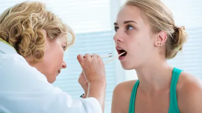
Because of this, you should still make an appointment with a doctor or dentist. They can assess the oral lesions and advise you on treatment or management options.
How is Leukoplakia Treated?
Treatment for leukoplakia often involves preventing the patches from becoming cancerous. These are most successful when started early while the patch is small.
Treatment may involve:
- Lifestyle changes — This involves maintaining a healthy diet, quitting tobacco and alcohol, taking supplements, or using mouthwash with ketorolac.
- Surgery — Although the surgeon can remove the lesions, there’s still a 10% to 20% chance that they’ll recur and a 3% to 12% chance of developing cancer in the area.
- Lasers — A surgeon can remove the lesions by using a high-energy laser.
- Electrocauterization — An alternative to laser removal. It involves using electrically heated instruments to remove oral leukoplakia patches.
- Cryotherapy — Involves targeting the lesions with liquid nitrogen to freeze and remove them.
- Photodynamic therapy — Treatment uses a light-sensitive medication and a light source to destroy abnormal tissue.
Your doctor will arrange regular follow-up visits with you as recurrences are common. Additionally, you usually don’t require treatment if you have hairy leukoplakia because it isn’t likely to lead to cancerous changes and cause no symptoms.
Instead, your doctor may recommend antiviral medications and topical treatments. Your doctor may also arrange follow-up visits to monitor your mouth or ongoing medication to prevent the patches from returning.
How to Prevent Leukoplakia?
As mentioned before a number of oral leukoplakia cases are related to smoking and alcohol. So, one of the easiest ways to prevent the condition is to avoid these products.
Good nutrition is another critical factor in preventing diseases and poor health. To reduce the likelihood of leukoplakia, follow a healthy, balanced diet rich in antioxidant foods. If you notice any changes in your mouth or think you have oral leukoplakia, contact your doctor or another healthcare provider for advice.
Leukoplakia: Symptoms, Causes and Treatment
NewMouth PodcastSources
- Mohammed, et al. “Oral Leukoplakia.” StatPearls Publishing, 2022
- Shang et al. “Association of Human Papillomavirus With Oral Lichen Planus and Oral Leukoplakia: A Meta-analysis.” Journal of Evidence Based Dental Practice, 2020
- Parlatescu et al. “Oral leukoplakia – an update.” Maedica, 2014
- Kusiak et al. “The Analysis of the Frequency of Leukoplakia in Reference of Tobacco Smoking among Northern Polish Population.” International Journal of Environmental Research and Public Health, 2020
- Shesha Prasad et al. “Idiopathic leukoplakia- report of a rare case and review.” J Clin Diagn Res, 2015.
- Alejandro. “New insights into the role of the oral leukoplakia microenvironment in malignant transformation.” Frontiers in Oral Health, 2024.

Licensed dental specialist focusing on personalized dental content writing and blogging.

Public health expert and copywriter covering various health topics, including dentistry.
Related Articles
6 Causes of Itchy Gums and How to Stop Them
Itchy gums are usually caused by plaque buildup, allergies, hormonal changes, dry mouth, or early gum disease. Learn wha...
Warts on the Tongue
Tongue Warts Warts are small, raised, fleshy bumps that can form anywhere on the body. They're usually harmless and pain...
Tonsillectomy: Purpose, Procedure, and Recovery
A tonsillectomy is a surgical procedure that removes the tonsils. Read this to learn more about tonsillectomy, its risks...

Gum Boils: Symptoms, Causes, Home Remedies & Treatments
What is a Gum Boil? A gum boil, or periodontal abscess, is a small, pus-filled bump on the gums. Often, it happens when...