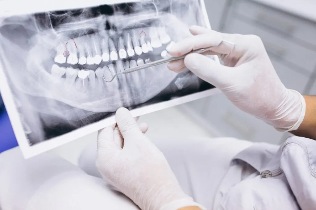
Dental X-rays: Types & Risks
Reader-supported. We may earn a commission from links on this page. Advertising disclosure.
In this article
Dental X-rays help find and treat dental issues early in development. Finding and treating oral problems early in their progression can help you:
- Improve the quality of your dental care
- Save money
- Prevent discomfort
- In extreme cases, save your life
Dental X-rays are often necessary if you need orthodontic treatment. General dentists and other healthcare providers use them to check the conditions of your mouth area. This includes:
- Teeth
- Teeth roots
- Jaw placement
- Facial bone structure
If you visit a new doctor, you’ll probably undergo a dental X-ray on your first visit. This helps them improve the quality of their dental practice and provide you with personalized oral care.
What are the Common Types of Dental X-rays?
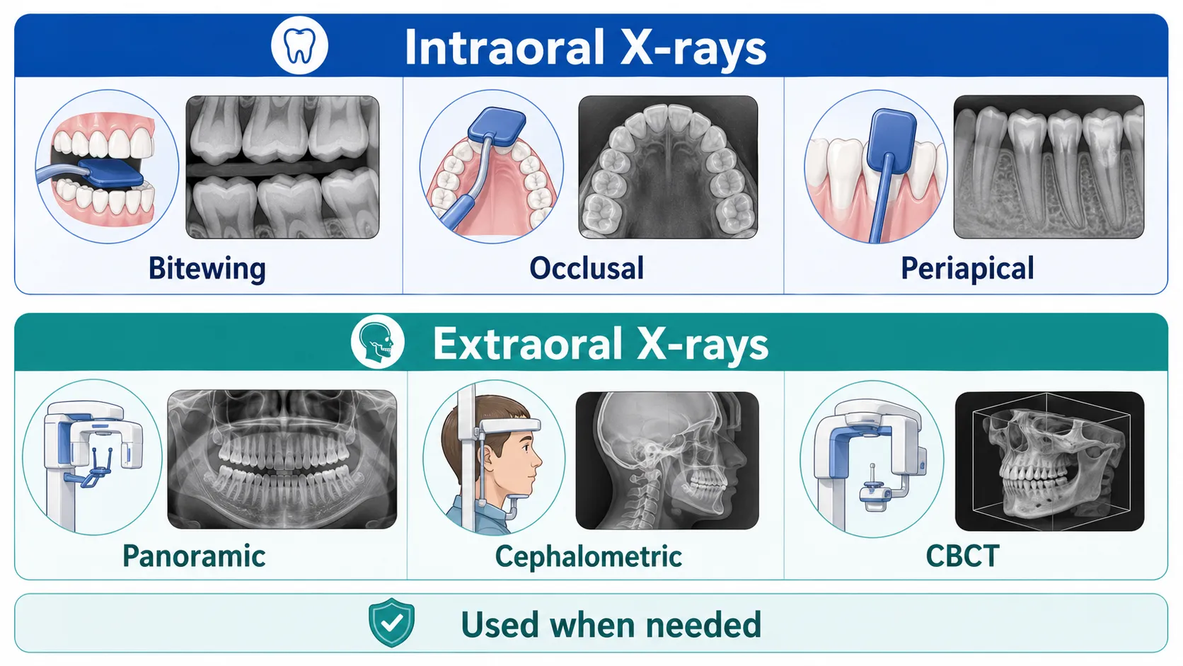
The two main types of X-rays in dentistry are intraoral and extraoral X-rays. Intraoral X-rays refer to when the X-ray film is in the mouth. Extraoral X-rays have a film outside the mouth.
What are Intraoral X-rays?
Intraoral X-rays are the more popular of the two types of dental X-rays. Intraoral X-rays offer much detail about the teeth.
Dentists use intraoral X-rays to do the following:
- Find cavities
- Assess the health of the tooth root
- Check the health of the bone surrounding the tooth
- Assess developing teeth
- Determine if a patient is at risk for periodontal disease
- Monitor the general oral health of the mouth
There are several types of intraoral X-rays, each displaying various aspects of the teeth:
1. Bitewing X-ray
Bitewing X-rays display details of the upper and lower teeth in a mouth area. Every bitewing scan shows a tooth from its crown to around the level of the supporting bone.
Bitewing X-rays detect tooth decay, bone loss, and adjustments in bone density due to gum disease. They also help assess the best fit of a crown or cast restoration.
2. Occlusal X-ray
Occlusal X-rays are more extensive scans that display full tooth development and placement. Every X-ray shows the complete arch of the teeth in either the upper or lower jaw.
Occlusal X-rays discover extra teeth, jaw fractures, a cleft palate, cysts, dental abscesses, or growths. They also find foreign objects lost in the mouth.
3. Periapical X-ray
Periapical X-rays reveal the entire tooth from the crown down to where the tooth anchors in the jaw. Every periapical X-ray displays this full tooth dimension.
It also shows all the teeth in one portion of the mouth, either in the upper or lower jaw. Periapical X-rays assess any teeth root abnormalities and the surrounding bone.
What are Extraoral X-rays?
Extraoral X-rays also show teeth. But unlike intraoral X-rays, they focus on the jaw and skull.
Extraoral X-rays don’t present in-depth details, so they don’t help find cavities or identify issues with individual teeth. Instead, dentists use extraoral X-rays for the following:
- Discover an impacted tooth or teeth
- Assess the growth and development of the jaw bones
- Identify issues between the teeth and the jaws
- Check for any problems relating to the facial bones
There are a few types of extraoral X-rays available, including:
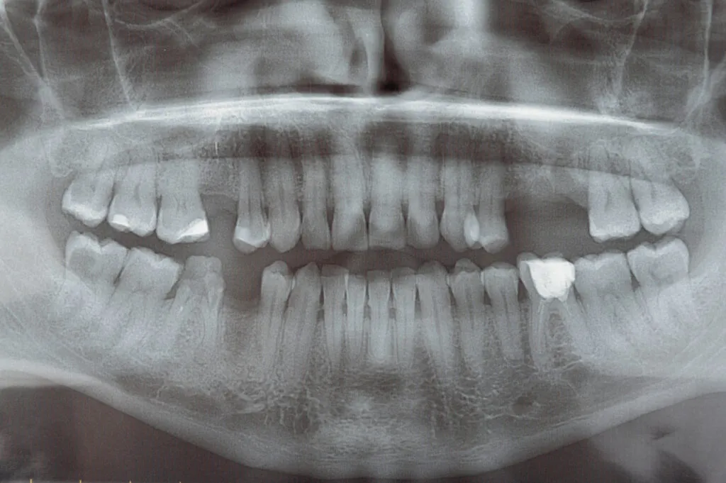
1. Panoramic X-ray
A panoramic X-ray reveals the whole mouth area. This type of X-ray identifies impacted teeth, shows your wisdom teeth’ progression, and helps diagnose tumors.
2. Tomogram X-ray
A tomogram X-ray displays a specific layer or ‘slice’ of the mouth while softening the focus on all other segments. This X-ray helps to assess structures that are difficult to observe.
3. Cephalometric X-ray
This type of X-ray displays the complete side of the head. Cephalometric X-rays examine the teeth concerning the jaw and your profile. Orthodontists commonly use it in diagnosing malocclusions.
4. Sialography X-ray
After injecting a special dye called a contrast medium into the salivary duct, a sialography X-ray scans the salivary glands. The pigment shows your salivary glands, soft tissues that aren’t normally visible on an X-ray. This procedure detects issues like blockages or Sjögren’s syndrome.
5. Computed Tomography X-ray
A computed tomography X-ray, or CT scanning, displays the body’s interior structure as a three-dimensional scan. It assesses issues related to the facial bones, including tumors and fractures.
6. Cone Beam Computed Tomography Imaging
CBCT shows a three-dimensional image of the face and jaws. It’s more common in dentistry than CT scanning.
Because it requires smaller, less expensive technology than an X-ray machine, many dentists offer CBCT imaging in their private practices. CBCT images are advantageous in planning dental implant placement, complicated extractions, and complex root canals.
How Often Should Dental X-rays Be Performed?
Doctors only perform dental X-rays when necessary. They’re not dangerous, but minimizing radiation exposure is always best. However, this varies by circumstance.
How often you should take X-rays depends on various factors, including:
- Medical and dental history
- Age
- Stage of the dental disease or condition
- Risk factors for different oral conditions
- Symptoms of oral disease
Tailored X-Ray Schedules
Some require dental X-rays every six months. Those with regular dental problems may need to take them more often. However, others with no history of dental or gum disease may only require a dental X-ray every few years.
Considerations for Children
Children should have fewer X-rays than adults because they’re less radiation-resistant. They shouldn’t have a dental X-ray more than once annually. However, once a child shows a higher risk for cavities, more X-rays may be necessary.
What are the Benefits of Dental X-Rays?
Dental X-rays are crucial in keeping your dental health in optimal condition. They provide valuable insights often not visible during a regular dental examination.
Some benefits of dental X-rays include:
Early Detection of Dental Issues
Dental X-rays can reveal problems that dentists can’t see with the naked eye. They’re beneficial in identifying dental issues at their earliest stages. This helps minimize the extent of damage so you can prevent costly and invasive procedures.
Full-Mouth Assessment
Dental X-rays provide dentists with a complete view of your mouth. They help assess and identify specific needs and risks. This leads to better and more precise treatment plans tailored to your unique oral health.
Treatment Planning
Dentists refer to Dental X-rays to plan for your treatment. The X-rays provide detailed images of the teeth, roots, and surrounding structures. This helps the dentist plan and execute the procedure more safely and accurately.
Documentation
X-rays serve as a record of your oral health over time. Dentists can use your regular dental X-rays as a baseline for comparison for future visits. It can help them track whether a procedure works or yields the desired changes.
What are the Complications & Risks of Dental X-rays?
Dental X-rays are very safe. While they work with radiation, the exposure levels are so low that they won’t harm children and adults.
However, like with any treatment, repeated exposure has some potential risks. These include:
- Thyroid cancer
- Tumors in tissues covering the brain and spinal cord
- Radiation exposure
- Affecting the health of developing fetus in pregnant women
How Can Your Dentist Minimize Radiation Exposure During Dental X-rays?
If you’re concerned, there are ways that your dentist can minimize the exposure to radiation from dental X-rays, including:
- Taking a single X-ray image rather than multiple
- Using the lowest radiation setting possible
- Protecting certain areas of the body from radiation using leaded coverings
How to Prepare for A Dental X-Ray?
Dental X-rays don’t require much preparation. All you need to do before meeting a dental professional is brush your teeth before the examination. This offers a cleaner and more hygienic space for the dentist to examine your mouth.
Summary
Dental X-rays are essential in diagnosing and preventing oral health issues. While radiation captures them, dental X-rays are safe, and the risks of harm are minimal. Receiving X-rays from professionals with proper experience will help guarantee your safety during the procedure.
Sources
- Mark, A. “Dental x-rays.” The Journal of the American Dental Association, 2019.
- Abrahams J.J. “Dental CT imaging: a look at the jaw.” Radiology, 2001.
- “The Selection of Patients for Dental Radiographic Examinations.” U.S. Food and Drug Administration, 2012.
- “New research on dental X-ray risks.” British Dental Journal, 2019.
- Hwang et al. “Health effects from exposure to dental diagnostic X-ray.” Toxicology and Environmental Health Sciences, 2018.
- Erdelyi et al. "Dental Diagnosis and Treatment Assessments: Between X-rays Radiography and Optical Coherence Tomography." Materials (Basel), 2020.
- “Dental x-rays.” University of Michigan School of Dentistry.

Experienced general dentist and adjunct professor. Advocates for preventive dentistry and dental education.

Freelance writer focused on authoritative dental content and oral hygiene guidance.
Related Articles

Teeth Cleaning Costs
Regular dental cleanings help prevent gum disease and tooth decay. Find out how much you'll pay for each type of cleanin...

Cost of Dental Check Ups
How Much Does a Dental Check-Up Cost? A dental checkup can cost between $0 to $500+ depending on several factors, includ...

Dental Anesthesia
Dental anesthesia eliminates pain and reduces anxiety during procedures. Here are the three types, their benefits, and p...

What is Gingivitis and How Do You Treat It?
Gingivitis can affect anyone at any age but is most common in adults. Learn what gingivitis is, its stages, the six symp...